Fig. 9.
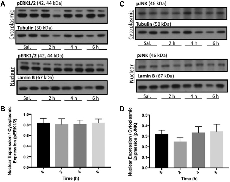
BMP-9 treatment does not activate mitogen-activated protein kinase signaling pathways. (A) Animals were administered a single dose of BMP-9 (1 μg/kg, i.p.) or 0.9% saline and treated for 2–6 hours. Following treatments, animals were sacrificed and brain microvessels were isolated and subjected to nuclear and cytoplasmic fractionation. Nuclear and cytoplasmic extracts were then prepared for western blot analysis. Fractionated microvessels (10 μg) were resolved on a 4%–12% SDS-polyacrylamide gel, transferred to polyvinylidene difluoride (PVDF) membrane and analyzed for expression of pERK1/2. (B) Relative levels of pERK1/2 expression in samples from A were determined by densitometric analysis and normalized to α-tubulin for cytoplasmic fraction and lamin B for nuclear fraction. (C) Fractionated microvessels (10 μg) were resolved on a 4%–12% SDS-polyacrylamide gel, transferred to PVDF membrane and analyzed for expression of phosphorylated JNK (pJNK). (D) Relative levels of pJNK expression in samples from (C) were determined by densitometric analysis and normalized to α-tubulin for cytoplasmic fraction and lamin B for nuclear fraction. Western blot data are reported as mean ± S.D. from at least three independent experiments, where each treatment group consisted of four individual animals (n = 4).

