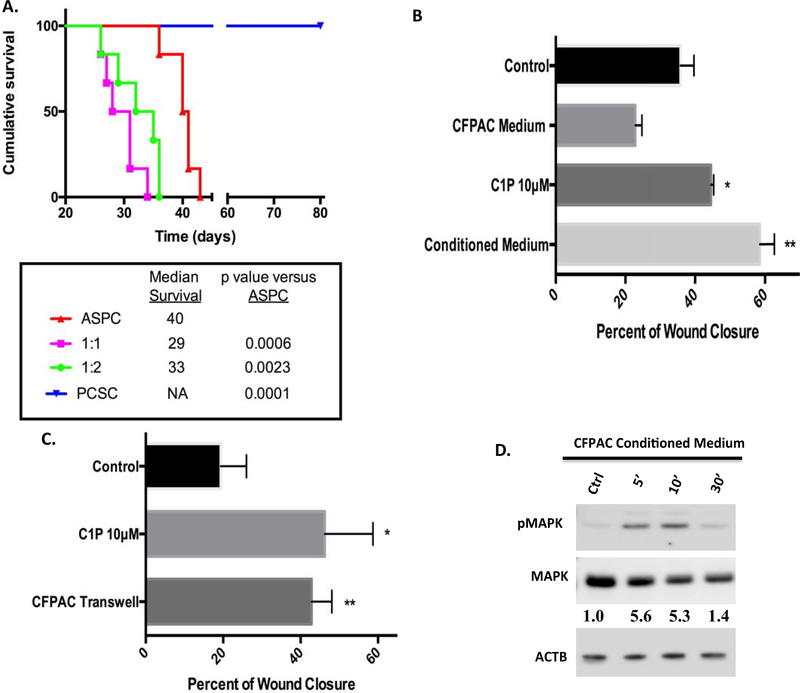
Figure 5:
CSCs co-injected with AsPC-1 cells reduces animal survival while CFPAC conditioned medium and CFPAC in transwells increased CSC migration. Studies using AsPC-1 cells (0.75 × 10⁶), AsPC-1 (0.75 × 10⁶), + CSC cells (0.75 × 10⁶), AsPC-1 (0.75 × 10⁶), + CSCs (1.5 × 10⁶), or CSCs alone (0.75 × 10⁶) were injected intraperitoneally into NOD/SCID mice. Survival from was noted from time of tumor cell injection. Statistical group differences in survival time were calculated using log rank testing (Mantel-Cox) with reduction in survival noted in combined injected cell populations PCSC / AsPC-1 (1:1- p=000.6 and 1:2- p =0.0023) as compared to AsPC-1 cells alone (A). Study was concluded on day 80 with no loss of CSC injected animals (p=0.0001, A) (n=6/group). Consistent with C1P enhanced migration (A, *p=0.0184, 45%± 0.43 SEM), CSCs treated with CFPAC-conditioned medium (B, **p=0.0025, 59%± 2.5 SEM) significantly increased wound closure in CSC scratch assays as compared to unconditioned CFPAC medium (23%± 1.1 SEM) or control medium (36%±2.2 SEM) (B, n=3/condition/experiment performed a minimum of 4 occasions). Transwell co-culture of CFPAC cells with wounded CSCs significantly enhanced wound closure (B,**p=0.471, 43%± 2.6 SEM) consistent with that seen with C1P (B,*p=0.0035, 46%± 6.2 SEM) as compared to control (C, 19%± 3.3 SEM) (n=4/condition/experiment performed a minimum of 3 occasions). CFPAC-conditioned medium induced a similar pattern of MAPK phosphorylation with densitometry analysis indicating a 5-fold increase in pMAPK (D, n=2,two different occasions, representative blot).

