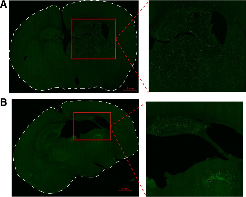
Fig. 2.

EGFP+ myeloid cell localisation in the brain after hypoxia-ischemia. a Representative tile-scanned confocal images of brain sections after hypoxia-ischemia (HI). a Dispersed pattern of EGFP+ myeloid cell infiltration in the hippocampus and thalamus 1 day after HI. b EGFP+ myeloid cells localised in the hippocampus and the white matter of the thalamus (medullary lamina of thalamus) in a spatially limited dense pattern 7 days after HI. n = 4/time point
