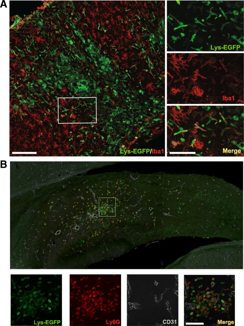
Fig. 3.

EGFP+ myeloid cells in the brain 7 days after hypoxia-ischemia are Iba-1− and Ly6G+. a EGFP+ cells are distinct from Iba-1-positive cells and display mostly round morphology in the ipsilateral cortex in animals with severe injury at 7 days after hypoxia-ischemia (HI). b Round-shaped EGFP+ cells are largely Ly6G+ and are not associated with CD31+ vessels (example from the hippocampus). Scale bars = 100 μm (at low magnification) and 50 μm (at high magnification)
