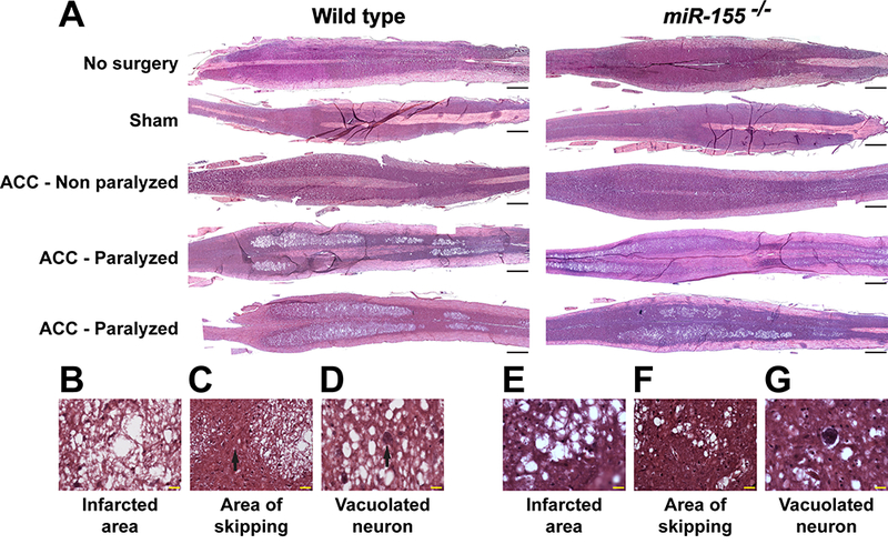
Fig. 2.

Correlation of spinal cord histological damage with paralysis in WT and miR-155−/− ACC mice. (A) Representative images of H&E-stained, longitudinal sections through the ventral horns of spinal cord of no-surgery, sham, ACC-Non-P and ACC-P WT and miR-155−/− mice, 44–48 h following ACC. Rostral is on the right side. Scale bars = 1 mm. (B, E) High definition of the large area of infarct from a WT (B) and a miR-155−/− (E) ACC-P mouse. Scale bars = 20 μm. (C, F) Skipping area [black arrow in (C)] in the same spinal cords as in (B) and (E), respectively, located between two infarcted areas. Scale bars = 20 μm. (D, G) Presence of vacuolated neurons in the same spinal cords as in (B) and (E), respectively. Scale bars = 20 μm.
