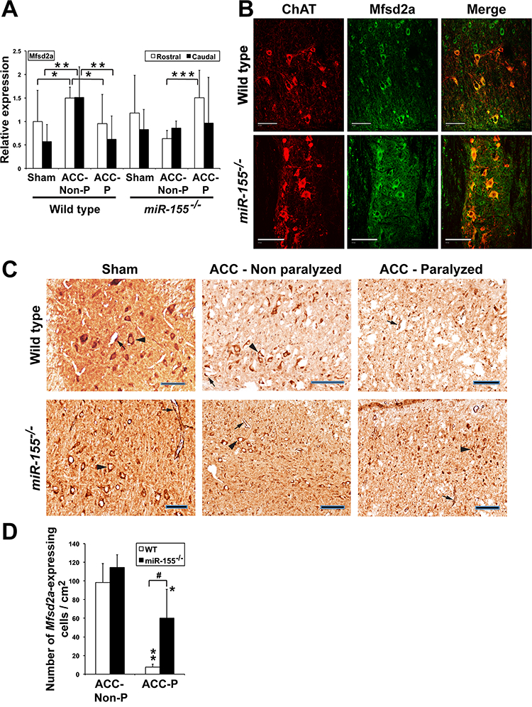
Fig. 4.

Mfsd2a expression is affected by ischemic conditions. (A) Mfsd2a relative expression (Mean + SD) 44–48 h after ACC in the rostral and caudal parts of spinal cord of WT sham (n = 9), ACC-Non-P (n = 5) and ACC-P (n = 7) mice, as well as of miR-155−/− sham (n = 5), ACC-Non-P (n = 5) and ACC-P (n = 10) mice, as shown by qRTPCR. *, P < 0.0668; **, P < 0.0361; ***, P < 0.00096. Values were normalized to WT rostral sham. (B) Mfsd2a (green) and ChAT (red), a marker of spinal cord moto-neurons, co-localize (yellow), in both sham WT and sham miR-155−/− mice, as shown by immunohistochemistry. Scale bars = 100 μm. (C) Mfsd2a-expressing cells in spinal cord of WT and miR-155−/− mice. Arrow heads: motoneurons. Arrows: endothelial cells. Scale bars = 100 μm. (D) Number of Mfsd2a-expressing endothelial cells and neurons (Mean + SD) 44–48 h after ACC in spinal cord of the same WT and miR-155−/− mice as in Fig. 3C (3 counts/ mouse). ** and *, ACC-P different from corresponding ACC-Non P; **, P < 0.000093; *, P < 0.0312;#,P < 0.0027. (For interpretation of the references to colour in this figure legend, the reader is referred to the web version of this article.)
