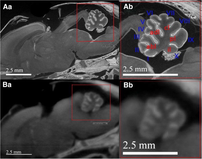
Fig. 5.
Direct comparison of ex vivo (spatial resolution of 10.7 μm/voxel) and in vivo (spatial resolution of 20 μm/voxel) micro-CT scans of the same 24-h-old rat’s head shows the image-quality difference between the two. (Aa) and (Ba) are sagittal illustrations of ex vivo and in vivo micro-CT scans, respectively. Gross neuroanatomy can be visualized in both scans with subtle difference appreciated by close-viewing, (Ab) and (Bb). Detailed cerebellar fissures, vermis (I–X), and lobes, can only be appreciated on ex vivo scans: abl = anterobasal lobe; adl = anterodorsal lobe; cl = central lobe; pl = posterior lobe; il = inferior lobe

