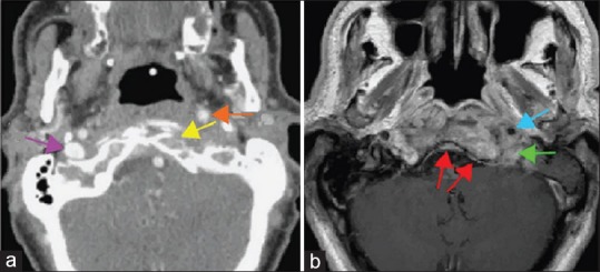
Figure 3.

(a) Contrast-enhanced computed tomography of the skull base shows erosion of the anterior cortex of the left occipital condyle (yellow arrows), anterior displacement of the left internal carotid artery (orange arrow). A normal right internal jugular vein is visible (purple arrow), whereas the left internal jugular vein is completely occluded. (b) Magnetic resonance imaging of the skull base shows pathological contrast enhancement in the clivus extending into the soft tissues surrounding the left internal carotid artery (blue arrow) and jugular foramen (green arrow). There is thickening and enhancement of the clival dura (red arrows)
