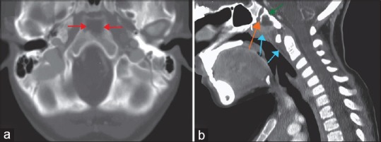
Figure 4.

A contrast-enhanced computed tomography scan (a) axial and (b) sagittal views in a child with pediatric clival osteomyelitis demonstrates destruction within the anterior aspect of the clivus bone (red and green arrows in a and b) posterior to the spheno-occipital synchondrosis (orange arrow) associated with anterior soft-tissue prominence extending close to the cephalic most portion of the adenoids along with some prevertebral soft-tissue thickening (blue arrows)
