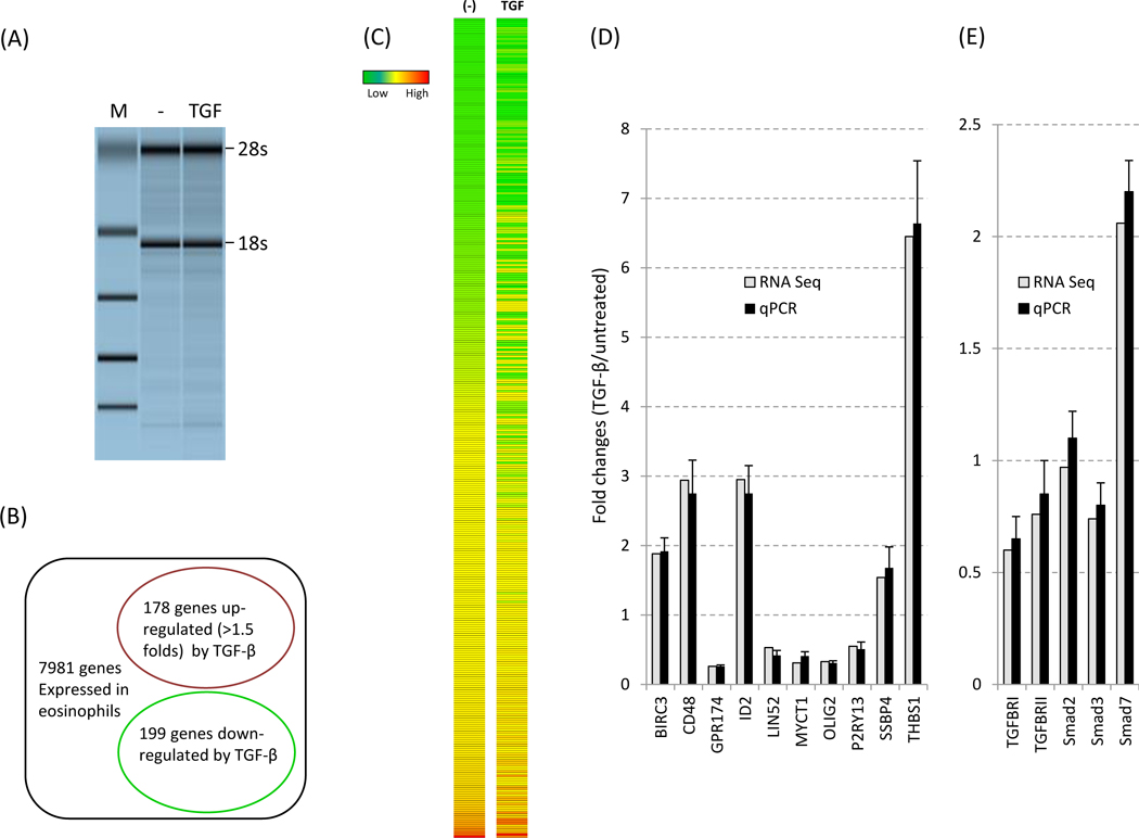
Figure 1. Expression of TGF-β target genes by human eosinophils.

(A) Cells were left untreated or treated with TGF-β (1ng/ml) for 5 h before harvest. RNA quality was determined as described in Methods before RNA-Seq anaysis. M: size marker. (B) Genes altered (cut off: 1.5 fold) by TGF-β are shown in the circles. The expression level of 7981 genes are greater than 2.00 (PKAM). (C) Heat map of the TGF-β responsive genes (377 genes total) is shown. (D) Validation of RNA-Seq by qPCR with the 10 select transcripts. qPCR data were obtained from 13 eosinophil donors and normalized to GAPDH expression. (E) Validation of RNA-Seq by qPCR with the 6 transcripts associated with canonical TGF-β signaling. Smad6 was undetectable (not shown). The fold change for each gene was calculated and compared between the two detection methods.
