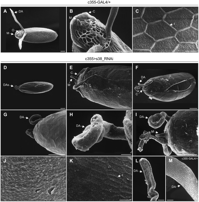
Figure 3.
Targeted downregulation of s38 protein in the ovarian follicle cells disrupts the patterning establishment of chorion’s regional specialization. (A–C) Scanning-electron-microscopy images of representative control (c355-GAL4/+) laid eggs. (A) Far-off view of the general egg morphology, featuring the two typically shaped and sized dorsal appendages. (B) The operculum region (orange-circled area) with the characteristic pattern of follicle-cell imprints on its surface and a morphologically undamaged micropyle. (C) High magnification scanning-electron-microscopy image of main-body egg’s surface, illustrating the characteristic hexagonal follicular imprints. (D–L) Scanning electron micrographs of representative s38-downregulated (c355 > s38_RNAi) laid eggs revealing diverse eggshell malformations. (D–G) Low magnification images of eggs being characterized by dysmorphic dorsal appendages with pleiotropic pathology, and variable sizes and shapes. (H,I) Close-up views of representative highly dysmorphic, thin and coiled dorsal appendages with abnormal tips. (J) The main-body surface morphology of an egg carrying short in length dorsal appendages is structured by a smooth and fragile eggshell without follicular imprints. (K,L) Eggs with longer but still abnormal in size dorsal appendages are presented with some follicular imprints in their main-body surface, albeit bearing much thinner ridges (K), while they encompass some typical features of physiological dorsal appendages (L). (M) A typical dorsal appendage with a paddle-shaped tip from a control laid egg, exhibiting discrete plaques on its dorsal surface. Number of eggs examined: control = 139, s38-downregulated = 271. DA: Dorsal Appendage(s), O: Operculum, M: Micropyle, C: Collar and I: Imprint. Scale Bars: 50 μm in (A,B,D–G and I) 10 μm in (C,H and J–M).

