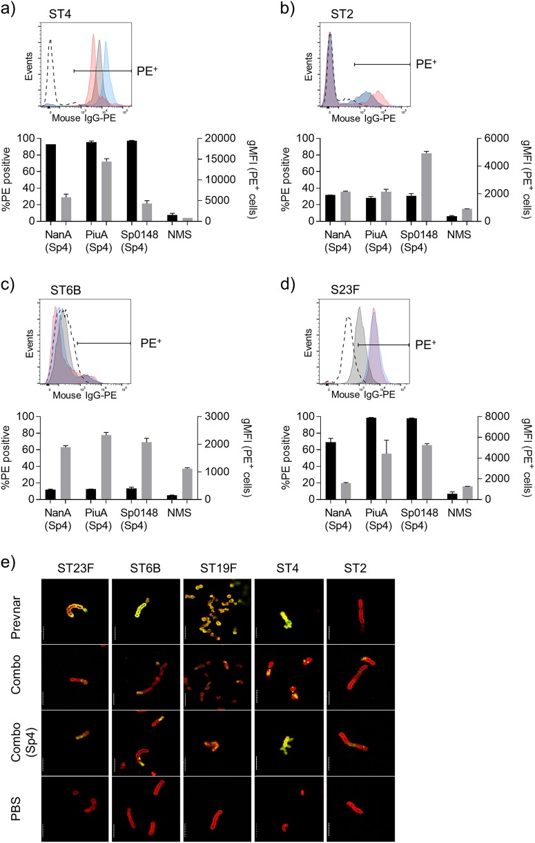
Fig. 4.
Antibody deposition on non-serotype 4 pneumococci. a-d Representative histograms and antibody deposition on homologous and heterologous pneumococcal isolates in 1% pooled antiserum from mice vaccinated with glycosylated NanA (grey shading), Sp0148 (red shading), or PiuA (blue shading) or normal mouse serum (dashed line). Black bars represent the percentage of PE+ bacteria and grey bars represent the gMFI of the positive population. Gates were set such that 5–10% of events were PE+ in the normal mouse serum (NMS) reactions to account for strain specific differences in auto fluorescence. Data are displayed as mean ± SEM from technical replicates. e Immunofluorescent staining of homologous and heterologous pneumococcal isolates using antiserum from mice vaccinated with the combination vaccine (green channel) and pneumococcal Omni serum (red channel). Length of scale bar is equal to 5 µm

