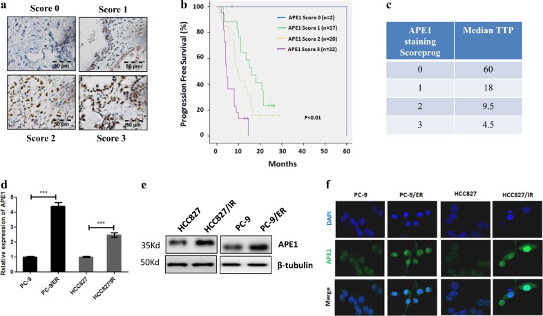
Fig. 1. The level of APE1 was closely associated with TKI resistance in LUAD.
a The expression of APE1 in LUAD specimens was measured by immunohistochemistry. b Kaplan–Meier analysis of progression-free survival rate for EGFR-mutated LUAD patients. Patients were divided into four groups based on APE1 staining score. c Median time to progress analysis. d qRT-PCR results show that the mRNA levels of APE1 were increased in TKI-resistant LUAD cell lines compared to their parental cell line. e Western blot analysis results show that protein levels of APE1 were increased in TKI-resistant LUAD cell lines compared to their parental cell line. f Immunofluorescence assay shows that protein levels of APE1 were increased in TKI-resistant LUAD cell lines compared to their parental cell line. HCC827/IR, Icotinib-resistant cell HCC827; PC-9/ER, Erlotinib-resistant cell PC-9; TTP time to progression; ***p < 0.001

