
Figure 5.
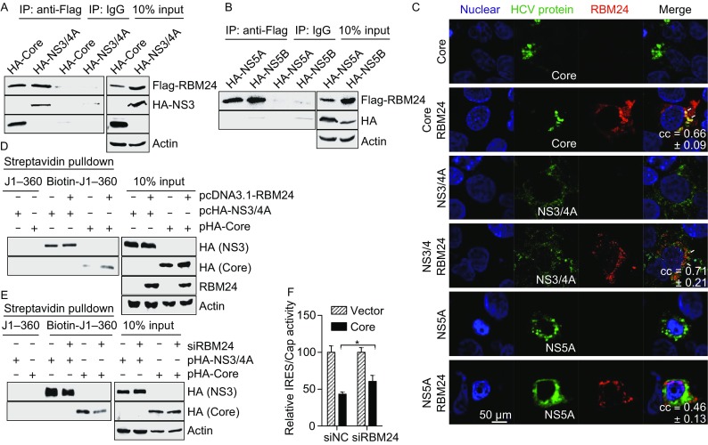
The interactions between RBM24 and HCV proteins. (A) 293T cells were transfected with Flag-RBM24 together with HA-Core or HA-NS3/4A. Cell lysates were immunoprecipitated with either an anti-HA mouse monoclonal antibody (HA) or a nonspecific mouse control antibody (IgG) in the presence of RNase A. Immunoprecipitated proteins were detected with an anti-HA rabbit monoclonal antibody (HA) or an anti-DYKDDDDK rabbit polyclonal antibody (Flag) correspondingly. (B) Co-IP was performed with the lysate of 293T cells transfected with pFlag-RBM24 and pHA-NS5A, or with pFlag-RBM24 and pHA-NS5B. (C) Huh7.5.1 cells were transfected with HCV protein (Core, NS3/4A or NS5A) expression plasmids together with pHA-RBM24. The localization of Core, NS3/4A, NS5A and RBM24 was detected by immunostaining with HA and Flag antibodies. The nuclei were stained with Hoechst 33258. (D) 293T cells were co-transfected with pcDNA3.1-RBM24 with either pHA-Core or pHA-NS3/4A. The cell lysates were incubated with the indicated biotin-labeled HCV fragments and affinity-precipitated with streptavidin beads. Precipitated proteins were detected by Western blot with the corresponding antibodies. (E) Huh7.5.1 cells were transfected with siRBM24 as described in experimental procedures and then transfected with either pHA-Core or pHA-NS3/4A. Cell lysates were incubated with the indicated biotin-labeled HCV fragments and affinity precipitated with streptavidin beads. Precipitated proteins were detected by WB with the corresponding antibodies. (F) Huh7.5.1 cells were transfected with indicated siRNAs first then transfected with pHCV-IRES and pHA-Core. Luciferase assay was performed 24 h later