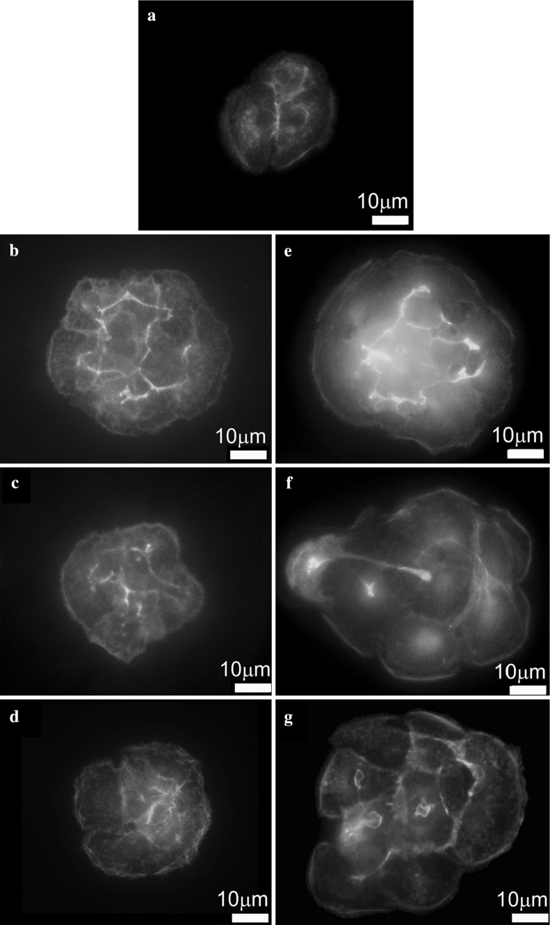
Fig. 4.
Fluorescence microscopy analysis of F-actin organization in HaCaT cells of: untreated cells (a); cells incubated with 0.5 μg/ml of P1 (b); cells incubated with 5 μg/ml of P1 (c); cells incubated with 50 μg/ml of P1 (d); cells incubated with 0.05 μg/ml of P2 (e); cells incubated with 0.5 μg/ml of P2 (f); cells incubated with 5 μg/ml of P2 (g)

