Fig. 6.
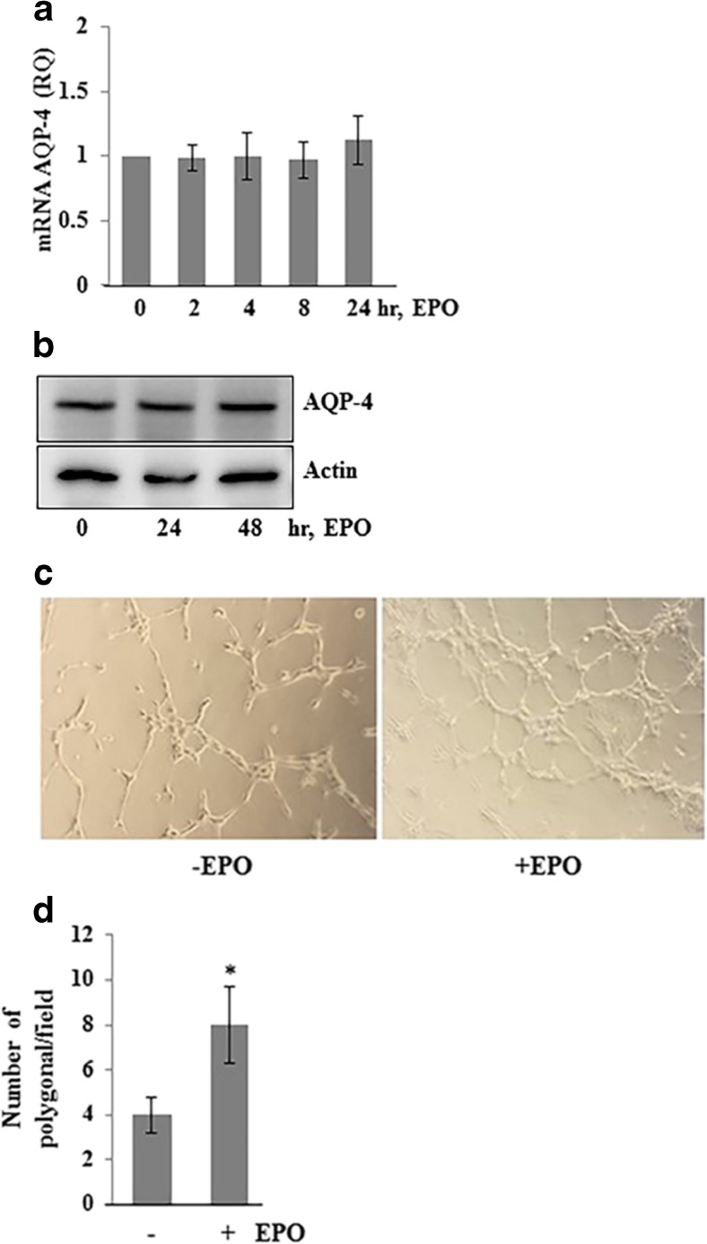
EPO-enhanced endothelial Matrigel tube formation network. (A) Confluent monolayer of HBME cells was treated with EPO (20 IU/ml) for 0, 2, 4, 8, and 24 h. Total RNA was isolated and AQP4 expression was analyzed with RT-PCR. (B) Confluent monolayer of HBME cells was treated with EPO (20 IU/ml) for 0, 24, and 48 h. Total protein was extracted and subjected to Western blotting to determine the AQP4 protein levels. The same blot was reprobed with anti-actin antibody for endogenous control. The blots are representative of three different experiments. (C) HBME cells were seeded on growth factor reduced Matrigel and treated without and with EPO (20 IU/ml) in serum-free, phenol red-free media. Representative images (× 20) show the network formed by HBME cells. (D) Polygonal area of network was counted in each field. The data shown here are from a representative of three experiments ± SEM. *p < 0.05

