
Figure 1.
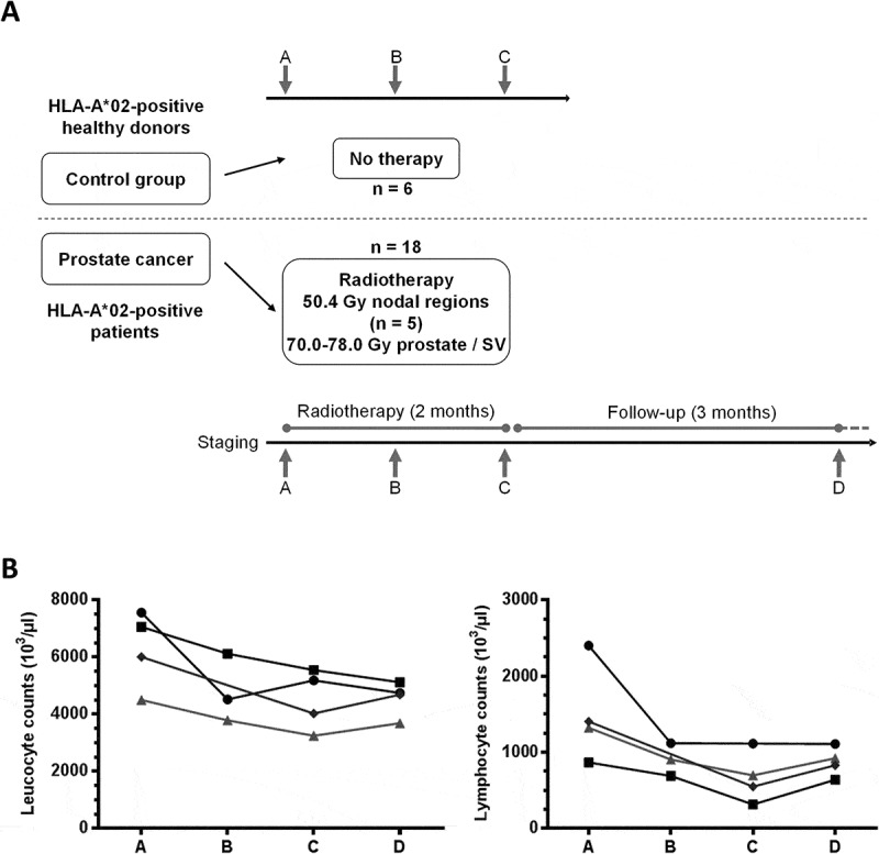
Flow chart of the study and white blood cells counts. (A) HLA-A*02+ patients undergoing primary RT for prostate cancer were included (n = 18). RT regimens are shown (SV = seminal vesicles). Blood samples were obtained before start of treatment (timepoint A), twice during therapy at 1 month intervals (timepoints B, C) and three months after the end of treatment at a follow-up visit (timepoint D). As controls, three consecutive blood samples (A, B, C) were obtained at one month intervals from HLA-A*02+ healthy donors (n = 6). (B) Absolute numbers of leucocytes (left) and lymphocytes (right) are shown before (A), during (B, C) and post-therapy (D) for four patients (four timepoints each, except for one patient for whom only 3 samples were available).