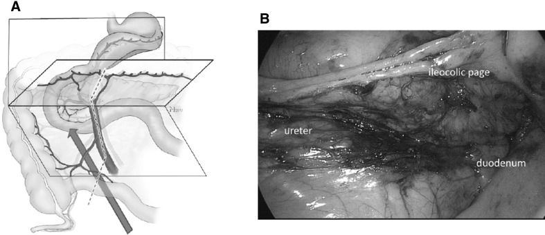
Fig. 2.
Step 1: the correct dissection plane between the retroperitoneum and the ileocolic page can be ensured when the duodenum is viewed from ventral and below (red arrow) (A). The mesenteric root (dotted line) must consequently be located ventral to the viewer´s eye. B shows a corresponding intraoperative view (see also Video No. 2) with the duodenum and pancreas dorsal and the mesocolon ventral to the level of dissection. This view from medial under the ileocolic page is established by the medial to lateral dissection approach exposing the retroperitoneum. (Color figure online)

