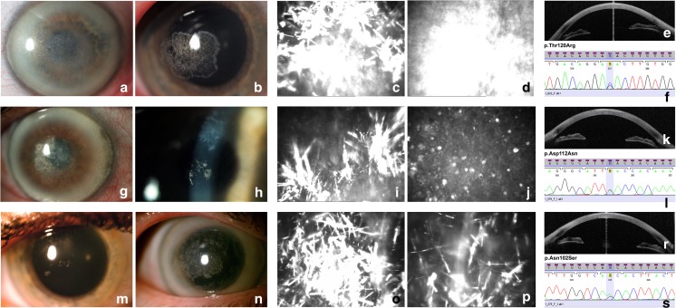
Fig. 2.
Corneal photographs and electropherograms of the corresponding UBIAD1 pathogenic variants. First two columns contain slit-lamp photographs showing crystalline formations (a, b, g, h, m, n) in the central and paracentral cornea, arcus lipoides (a, g, n), and haze (a, g, n). Columns three and four contain IVCM images presenting spindle-shaped corneal deposits (c, i, o, p), homogeneous conglomerate of deposits (d), and microcysts at the epithelial level (j). The last column includes AS-OCT images with sagittal sections demonstrating hyperreflective opacities in the anterior part of the corneal stroma (e, k, r). Electropherograms from Sanger sequencing of UBIAD1 exon 1 showing the identified c.359C > G (p.Thr120Arg) (f), c.334G > A (p.Asp112Asn) (l), and c.305A > G (p.Asn102Ser) (s) pathogenic variants

