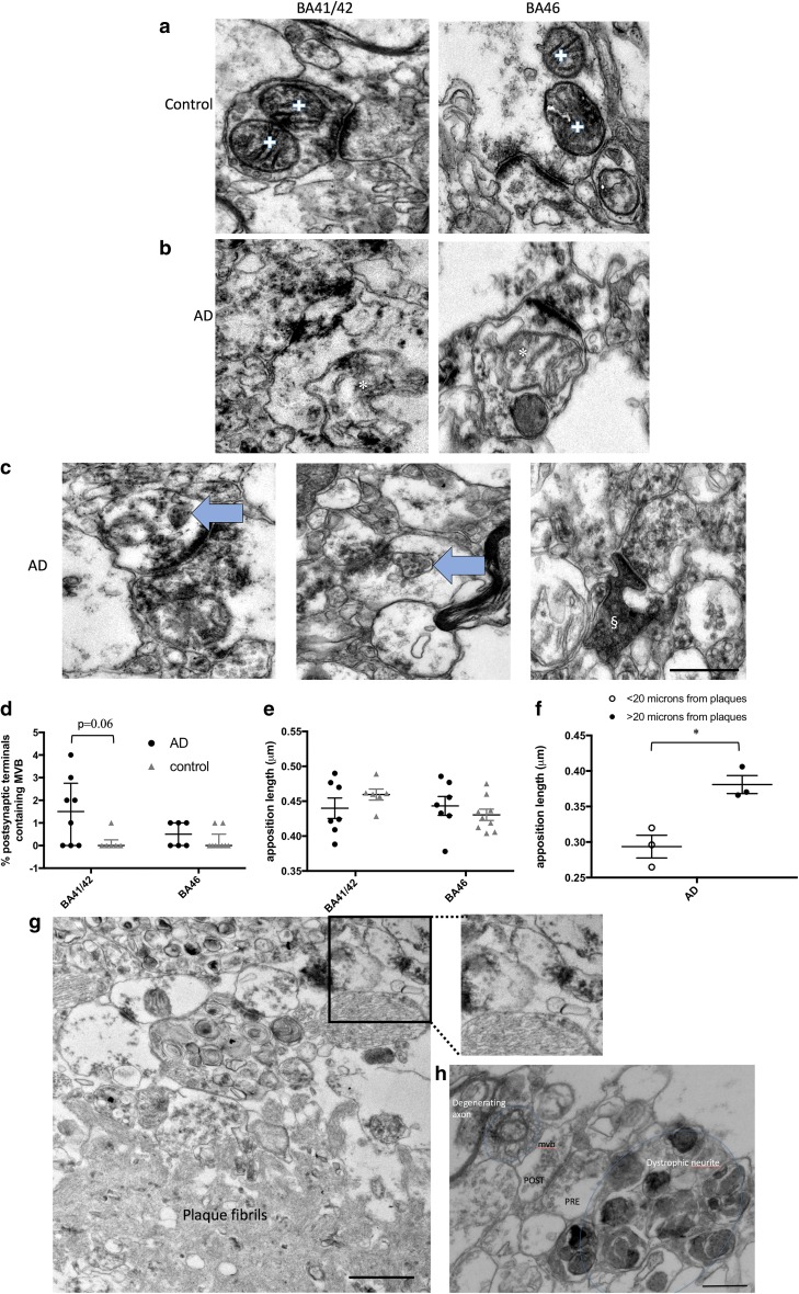
Fig. 4.
Changes in synaptic morphology in AD. In control cases, multiple mitochondrial profiles in individual pre-synapses were observed (a, crosses), while in AD cases, mitochondria with irregular profiles were observed in synapses (b, asterisks). Multivesicular bodies (MVB, arrows, c) were observed in a subset of post-synapses, and occasional dark degenerating spines (§, c) were observed in AD cortex. MVB appeared most often in AD BA41/42 synapses where there was a trend to increase compared to control (d, Kruskal–Wallis, p = 0.06). Apposition length was unchanged in AD vs. controls in BA41/42 or BA46 (e). When more blocks were examined from temporal, frontal and occipital regions to find synapses near plaques (f, g) and the data combined, we observe significantly decreased apposition length in synapses near plaques compared to those far from plaques (f, asterisk: unpaired t test with Welch correction, t = 4.28, p = 0.01). g An example of a small synapse near a plaque. h More detail around a plaque including a synapses (with pre- and post-synaptic terminals labelled, the post-synapse contains a MVB), a dystrophic neurite, and a degenerating axon. Data are shown as median with interquartile range. Scale bars represent 500 nm (a–c, inset g, h); 1000 nm (large panel g)

