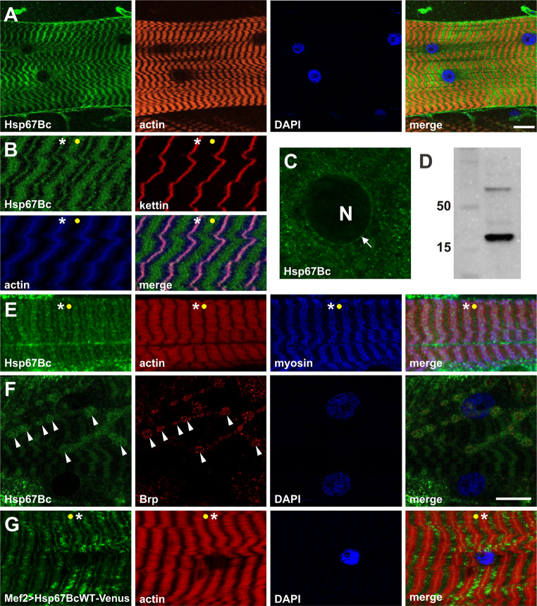
Fig. 2.
Expression pattern of Hsp67Bc in larval body wall muscles. a Hsp67Bc displays sarcoplasmic-associated expression in third instar larval muscles. Longitudinal optical sections of ventral longitudinal VL4. Anti-Hsp67Bc [8], TRITC–phalloidin for F-actin, DAPI. Scale bar: 15 μm. b Sarcomeric localization of Hsp67Bc. It is detected at the Z-bands (white asterisks) and in A-bands (yellow dot). Magnified longitudinal optical sections of ventral longitudinal VL4. Anti-Hsp67Bc [8], anti-kettin (DSHB), TRITC–phalloidin for F-actin, DAPI. c Hsp67Bc accumulation at the external surface of the nucleus (white arrow), N indicates nucleus. Magnified longitudinal optical sections of ventral longitudinal VL4. Anti-Hsp67Bc [8]. d Western blot of the third instar larvae protein extract showing the 22 kDa band of the expected size of the Hsp67Bc protein. e Sarcomeric localization of Hsp67Bc. Hsp67Bc is detected at the Z-bands (white asterisks) and in A-bands (yellow dot). Magnified longitudinal optical sections of ventral longitudinal VL4. Anti-Hsp67Bc [8], anti-MHC (gift from D. Kiehart),TRITC–phalloidin for F-actin, DAPI. f Sarcoplasmic distribution of Hsp67Bc. A small fraction of Hsp67Bc localizes at the NMJ (white arrowheads). Longitudinal optical sections of lateral longitudinal LL1. Anti-Hsp67Bc [8], anti-Brp1 (DSHB), DAPI. Scale bar: 15 μm. g Sarcomeric localization of Hsp67BcWT-Venus in Mef2 > Hsp67BcWT-Venus larvae. Like endogenous Hsp67Bc, it is detected at the Z-bands (white asterisks) and in A-bands (yellow dot). Local accumulations of overexpressed protein did not lead to destabilization of the myofibrillar organization. Magnified longitudinal optical sections of ventral longitudinal VL4. TRITC–phalloidin for F-actin, DAPI

