Fig. 5.
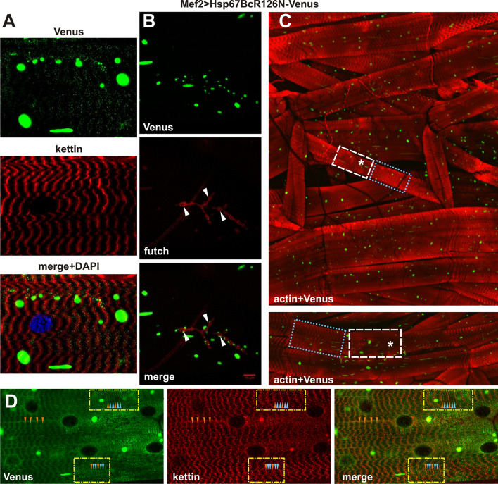
Phenotypic analysis of Mef2 > Hsp67BcR126N-Venus larval muscles. a Hsp67BcR126N-Venus distribution is disrupted to a significantly lesser extent. Longitudinal optical sections of ventral longitudinal VL4. Note the presence of large green aggregates. Anti-kettin (DSHB), DAPI. b The aggregate-prone form of Hsp67BcR126N-Venus retained its ability to localize at the NMJ sites (white arrowheads). Note the presence of large green aggregates. Anti-futsch/22C10 (DSHB). Scale bar: 15 μm. c Hsp67BcR126N-Venus distribution in ventral part of hemisegment musculature. Besides regions which appeared normal with regularly interspaced Z-bands (blue dotted box), we observed neighboring regions with attenuated intensity of actin bands (asterisks) and regions with a condensed sarcomeric pattern (white dashed box). Note the presence of large green aggregates. Inset in the bottom shows magnified longitudinal optical sections of ventral longitudinals VL4 and VL3. 3D reconstruction of confocal scans through ventral part of hemisegment musculature (Olympus FV1000 software). TRITC–phalloidin for F-actin. d Magnified longitudinal optical sections of ventral longitudinal VL4. Hsp67BcR126N-Venus localized in additional stripes seen as two bands (blue arrowheads) on each side of the Z-line (orange arrowheads). Note the presence of large green aggregates. Anti-kettin (DSHB), DAPI

