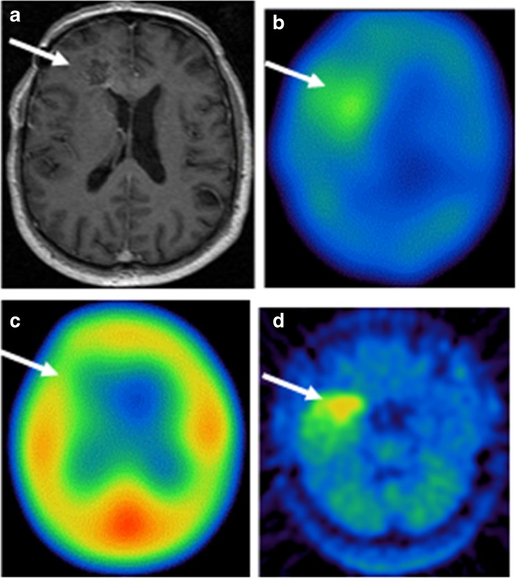
Fig. 1.
A 66-year-old man with grade IV glioma in the right frontal lobe. Axial T1-weighted MRI showed the lesion in the right frontal lobe with enhancement (a). The tumor lesion was positive in [123I]-VEGF SPECT imaging 18 h after injection of [123I]-VEGF (b), and no significant [123I]-VEGF accumulation was demonstrated 30 min after the [123I]-VEGF application (c). The lesion was positive in [11C]-MET PET (d). White arrows indicate the tumor region

