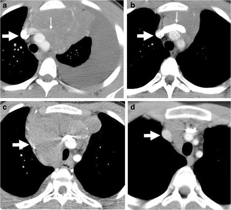
Fig. 6.
Vascular compression by mediastinal mass. a Pre-study axial CT image in a 7-year-old boy shows the contrast-enhanced left innominate vein is obliterated (thin arrow); (b) post-therapy, axial CT shows the caliber of the left innominate vein has been reconstituted. The SVC in both images of this boy (broad arrow) is patent. c, d Axial CT images in a 14-year-old boy show almost total compression of the SVC laterally on the pre-therapy study (c, arrow), which returns to normal caliber as seen in (d) post-therapy. SVC superior vena cava

