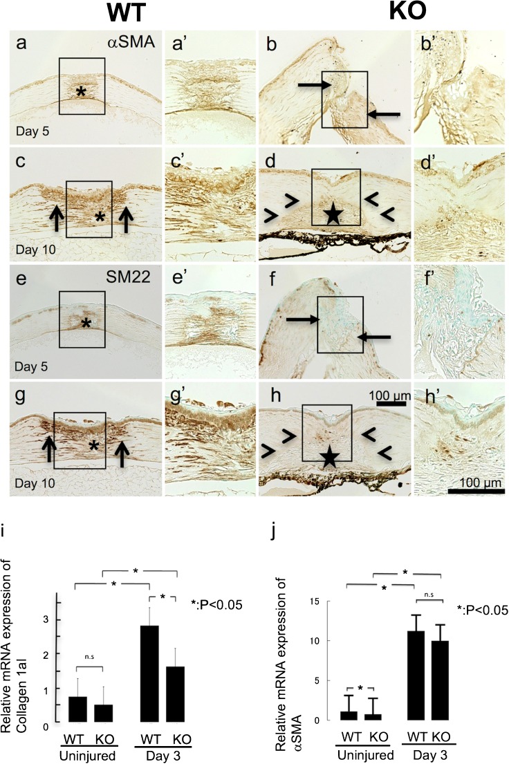
Fig. 3.
Detection of myofibroblasts in a healing incision-injured cornea of a wild-type (WT) or a TRPV1-null (KO) mouse based on gene expression analysis on collagen 1a1 and α-smooth muscle actin (αSMA) and immunohistochemistry for αSMA and SM22 in healing corneas
At day 5, the healing granulation tissue formed in the wound is occupied with αSMA-labeled myofibroblasts (asterisk) in a WT cornea (a). On the other hand, few myofibroblast is observed in a KO stroma, even in the area adjacent to the incision (arrows) (b). At day 10, myofibroblasts are distributed not only in the scar tissue (asterisk) but also in the stroma adjacent to the incision injury (arrows) in a WT tissue (c). Myofibroblasts are also seen in the granulation tissue (star) formed in the incision in a KO stroma, but the immunoreactivity seems less intense in a KO tissue as compared with a WT cornea (d). It is to be noted that the stroma adjacent to the granulation tissue is free from myofibroblasts (arrowheads) in a KO corneal stroma (d). Panels (e–h) show the expression pattern of SM22, another marker for myofibroblast. SM22 expression pattern is similar to that for αSMA indicated in panels (a–d). mRNA expression of collagen 1a1 is upregulated by the injury at day 3 as compared with baseline expression level (i). Such upregulation is counteracted by the loss of TRPV1. mRNA expression of αSMA is suppressed in an uninjured cornea by TRPV1 gene ablation. However, upregulation of αSMA is not affected by TRPV1 knockout at day 3 (j)

