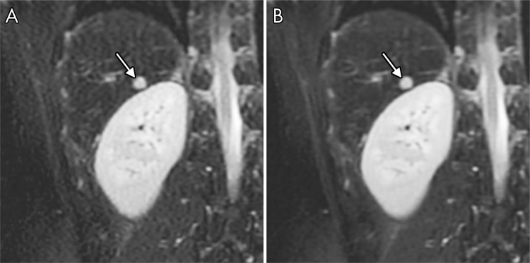
Figure 5:
Non–contrast-enhanced coronal images of liver lesion (arrow) in a 19-year-old man reconstructed with, A, conventional parallel imaging and compressed sensing (PICS) reconstruction and, B, variational network (VN) approach. VN approach achieved comparable images of lesion with improved perceived SNR.

