
Figure 2. ABCA1 increases V-ATPase on the cell surface.
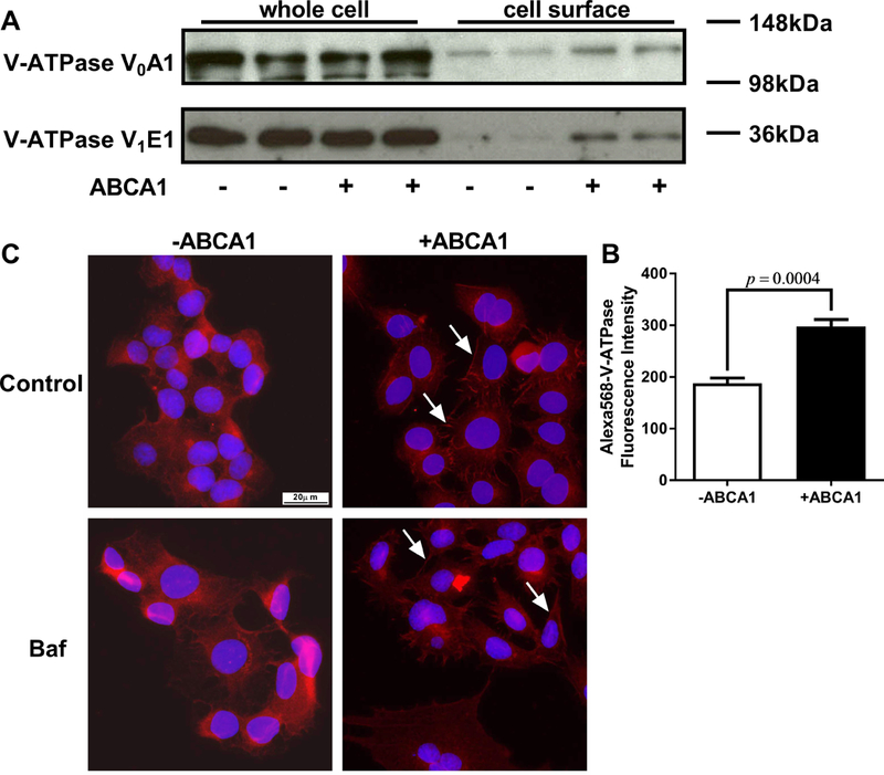
A. Cell surface proteins from ABCA1 induced and non-induced BHK cells were biotinylated, treated with the cell permeable cross linker DSP, and separated from the cellular lysate by streptavidin pull down. Total and cell surface V-ATPase subunits were detected by Western blot analysis. B. ABCA1 induced and non-induced BHK cells were fixed without permeabilization and were stained with antibody against V-ATPase V0A1. Flow cytometry was used to measure cell surface V-ATPase (N=3; mean ± SD; P=0.0004). C. ABCA1 induced and non-induced BHK cells were pretreated +/−10 μM bafilomycin A1 for 1 hr, fixed, permeabilized, and stained with antibody against V-ATPase V0A1 followed by epifluorescent microscopy (40× objective) for V-ATPase (red) and DAPI (blue). ABCA1 expression increased the level of V-ATPase on the cell surface (white arrows) independent of bafilomycin A1 treatment.