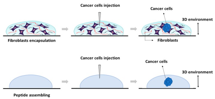
Figure 5.
Schematic representation of the 3D system’s construction using the self-assembling peptide scaffold RAD16-I. The system was developed (from left to right) as follows: First, peptide was assembled in presence (top) or absence of fibroblasts (bottom). Then, cancer cells were injected into each preformed matrix (center). The resulting 3D cultures were cancer cells embedded into a fibroblasts matrix (top) or a cancer cell into an empty peptide gel (bottom).

