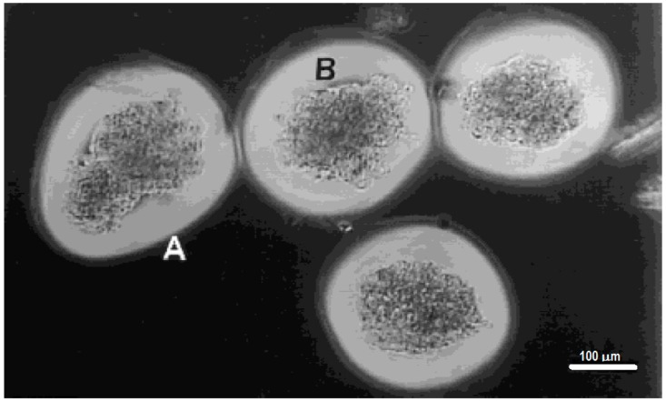
Figure 5.
Porcine islet cells encapsulated with a PEG diacrylate-based hydrogel using interfacial photopolymerization. The thin, outermost zone (labeled A) was presumed to be less crosslinked, and the filamentous nature of this outer zone may be due to the incomplete crosslinking of the hydrogel at the termination of laser illumination. Because the photoinitiator was present only at the islet surface, the thicker inner zone, closer to the islets (labeled B) and thus closer to the eosin Y photoinitiator, was presumably more highly crosslinking and thus had a more dense appearance. (Reproduced and adapted with permission from [20].)

