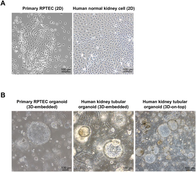
Fig 1. Primary renal proximal tubular epithelial cells (RPTECs) and normal human kidney cells form tubulocysts.
(A) Phase-contrast images: primary RPTECs and normal human kidney cells showing similar cellular morphologies in 2D cultures. (B) Phase-contrast images: spherule formation observed in primary RPTECs and normal human kidney cells. The 3D-on-top assay provides a clearer image of human normal kidney cell organoids than the 3D-embedded assay.

