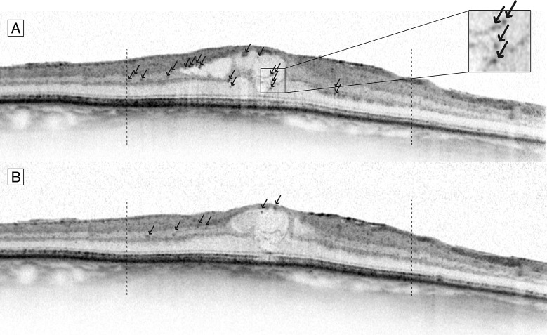
Fig 1. Hyperreflective foci on SD-OCT before and after treatment with anti-VEGF.
Foveal centered spectral domain optical coherence tomography (SD-OCT) B-scan image of a patient with DME before (A) and after (B) 3 injections with anti-VEGF. Black arrows indicate hyperreflective foci, within 3000 μm of the fovea (dashed bars).

