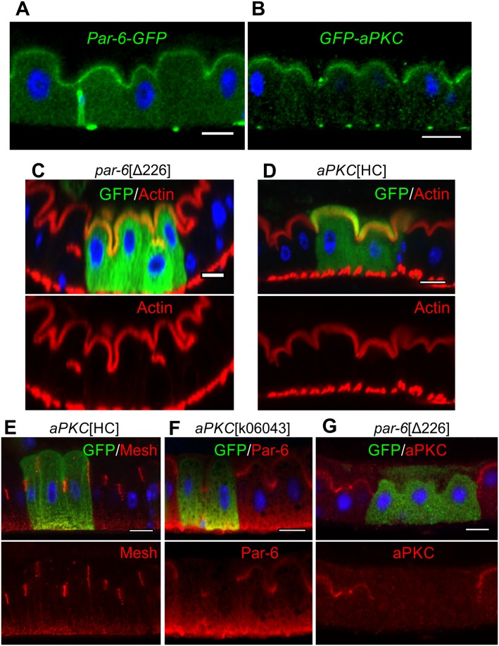
Fig 3. aPKC and Par-6 are not required for EC polarity.
Subcellular localisation of endogenously tagged Par-6–GFP (A) and GFP-aPKC (B), as revealed by anti-GFP staining (green). MARCM clones (marked by GFP, green) homozygous mutant for par-6Δ226 (C) and aPKCHC (D, E) show normal apical actin brush borders (F-actin in red) and SJ localisation (E; Mesh, red). The apical localisation of Par-6 (red) is lost in aPKCK06043 MARCM clones (marked by GFP, green) (F) as is the apical localisation of aPKC (red) in par6Δ226 MARCM clones (marked by GFP, green) (G). Scale bars, 10 μm. aPKC, atypical protein kinase C; EC, enterocyte; GFP, green fluorescent protein; MARCM, mosaic analysis with a repressible cell marker; SJ, septate junction.

