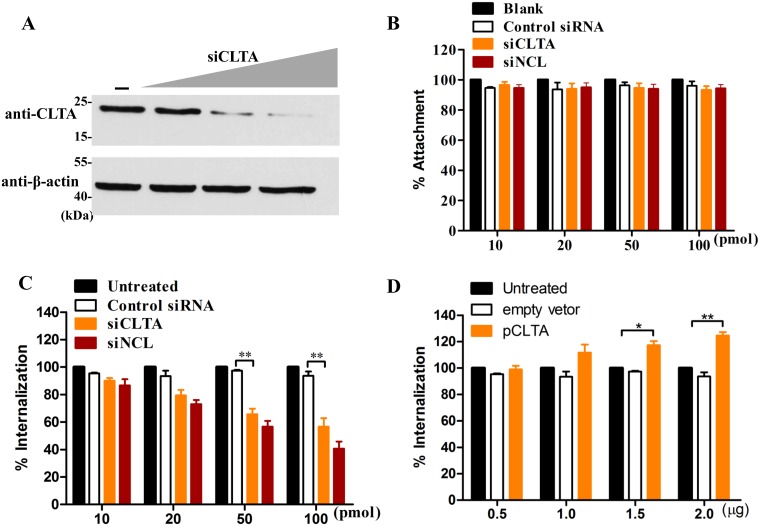
Fig 8. The interaction between CLTA and NCL plays an important role in RHDV internalization.
(A) The expression level of CLTA was determined by western blot analysis with anti-CLTA mAb. Cell lysates were obtained from RK-13 cells transfected with CLTA siRNA (10, 20, 50, or 100 nM) for 24 h. β-actin was employed as an internal control. (B) The effect of CLTA siRNA on RHDV attachment. Before the addition of RHDV (MOI = 1) for 2 h at 4°C, RK-13 cells were transfected with CLTA siRNA. Non-specific siRNA was used as a control. siNCL acted as a positive control. The percentage of attached RHDV VP60 copies was calculated as a ratio to the value obtained from untreated RK-13 cells. (C-D) The effect of CLTA on RHDV internalization. Before the addition of RHDV (MOI = 1) for 2 h at 4°C, RK-13 cells were transfected with CLTA siRNA (C) or the pCLTA plasmid (D). Non-specific siRNA or pCMV-Myc was used as negative control. siNCL acted as a positive control. The percentage of internalized VP60 copies was calculated as a ratio to the value obtained from untreated RK-13 cells. The student’s t-tests and analyses of variance were used for statistical analyses, *p < 0.05 and **p < 0.01. The experiments were conducted in triplicate and produced similar results.

