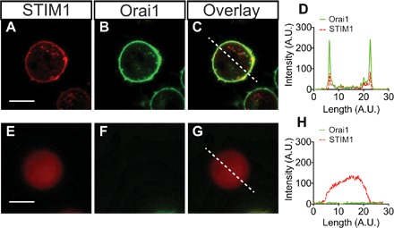
Fig. 6. STIM1 peptide interaction with the cell membrane protein Orai1 in HEK 293 cells.

(A) GFP-tagged Orai1 is present only at the cell membrane surface. (B and C) After delivery, Alexa Fluor 647–tagged STIM1 localizes to the cell surface, with a strong overlap with Orai1 (C), indicating that specific binding has occurred. (D) A line trace through the composite image (dashed line) shows colocalization of the STIM1 and Orai1 proteins at the membrane. (E to H) In Orai1-negative cells (E), the STIM1 is uniformly distributed throughout the cytosol. Scale bar, 5 μm. (H) The composite cross section shows that STIM1 does not localize to the cell membrane without the presence of Orai1.
