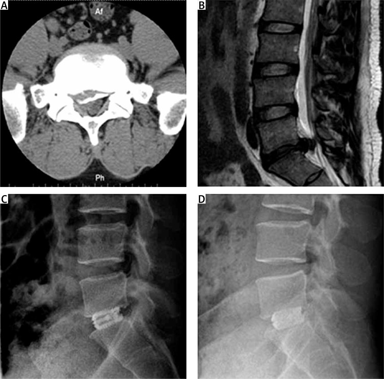
Figure 1.
Pre- and post-operative images of a patient who received SAEFC. A – The preoperative image of CT scan for central disc herniation, which was accompanied by calcification. B – Preoperative T2 WI MRI scanning for protrusion of intervertebral disc at L5/S1. C – X-ray image of lateral projection detected on 1 week after operation. D – X-ray image of lateral projection detected 1 year after operation

