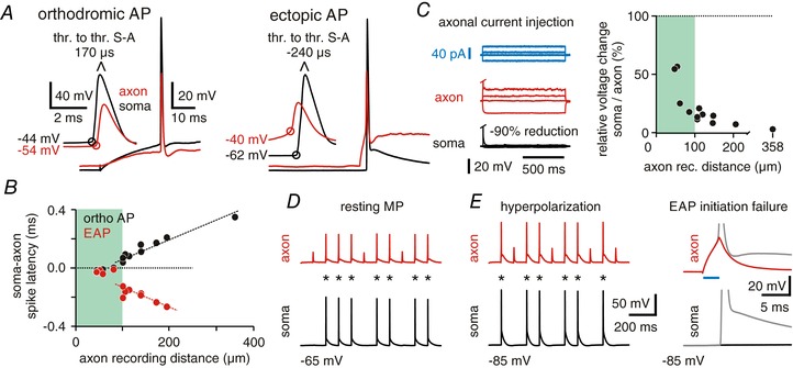
Figure 2. Ectopic action potentials reliably propagate back to the somatic compartment.

A, characteristics of orthodromic and ectopic action potentials (EAPs). Left panel: action potentials (APs) triggered by somatic current occur first in the soma and after a short delay at the axonal recording pipette; right panel: EAPs are first recorded in the axon and then at the somatic recording site where they arise directly from resting membrane potential. Both recordings are from the same cell, distance between somatic and axonal recording was 181 μm (voltage values indicate AP thresholds; resting membrane potential at soma and axon was −63 mV; thr. to thr.: interval between AP thresholds). B, scatter plot depicts latencies between somatic and axonal recordings of orthodromic and ectopic APs for all recordings. Note the near simultaneous occurrence of orthodromic APs at proximal recording distances, in line with spike initiation at the distal AIS. C, long current pulses (1 s) were injected into the axon through the patch pipette (blue trace). Resulting voltage deflections are very small in the soma (black trace, median = −85.6%, 13 cells) compared to the axon (red). Right panel: ratio of somatic to axonal voltage deflections vs. axonal recording distance. D, all EAPs evoked in the axon (red) reliably propagate back to the soma (black) at resting membrane potential (n = 2355 EAPs, 13 cells). Asterisks mark EAP induction. E, hyperpolarization of the soma induces no propagation failures (9 out of 10 cells). Asterisks mark EAP detection. Right: traces of EAPs (grey) and initiation failures taken from traces on the left (blue: duration of axonal current injection).
