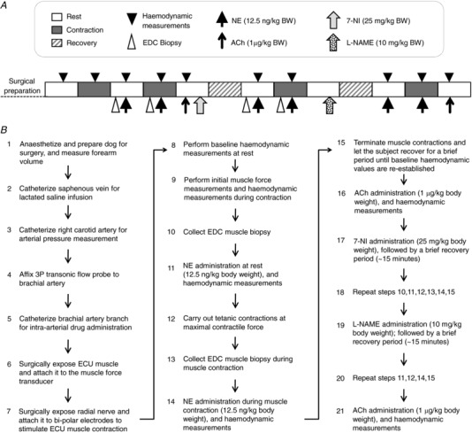
Figure 1. A schematic outline of the experimental protocol for studying sympatholysis in dogs.

A, experimental steps are shown from left to right starting from surgical preparation of the subject. Boxes show the status of muscle (rest, contraction or recovery). B, a flow chart of the experimental protocol. BW, body weight; ECU, extensor carpi ulnaris muscle; EDC, extensor digitorum communis muscle.
