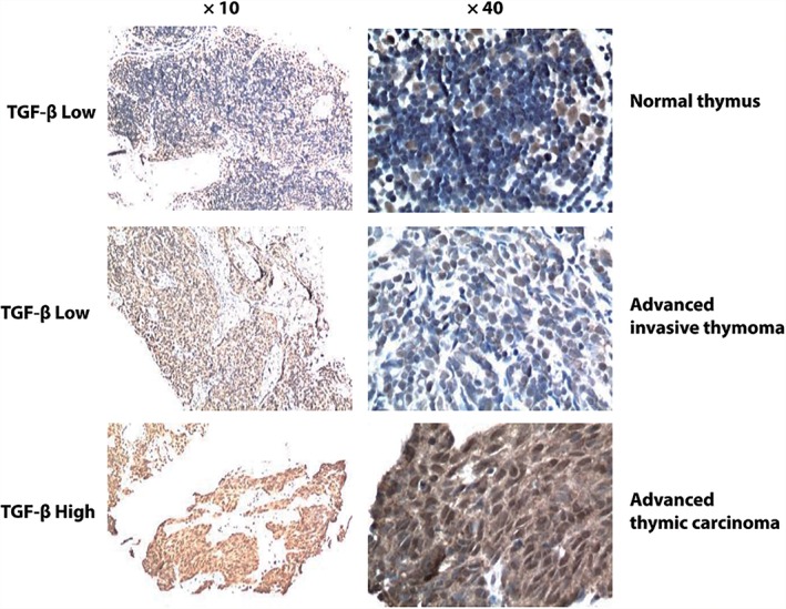
Figure 2.

Representative images of transforming growth factor‐β (TGF‐β) staining in normal thymus tissue and advanced thymic epithelial tumor (TET) pre‐treatment specimens. The median TGF‐β immunohistochemistry expression score is 4. High TGF‐β expression was identified with a score > 4, while low expression was ≤ 4. Basal level TGF‐β staining could be observed in the non‐thymocytes located in the medulla of normal thymus tissue.
