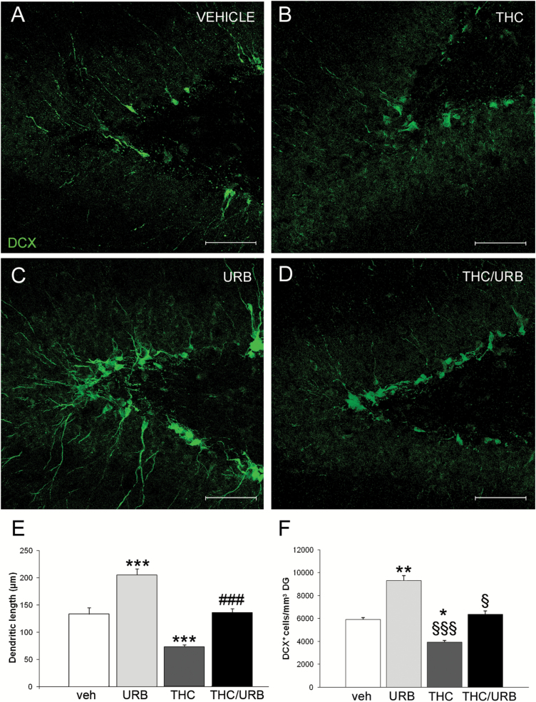
Figure 3.
(A-D) Representative confocal microscopy images of immunofluorescent doublecortin (DCX) labeling (green) in dentate gyrus (DG) of vehicle-, Δ9-tetrahydrocannabinol (THC)-, URB-, and THC/URB-treated rats. Scale bar: 75 µm. (E) Quantification of apical dendrite length in DG DCX+ neuroblasts. Compared with the vehicle-treated group, DCX+ cells had significantly longer apical dendrites in URB-treated rats. Conversely, mean dendritic length of DCX+ cells was significantly decreased in THC- vs vehicle-treated rats. No difference was observed between vehicle- and THC/URB-treated groups. Data are expressed as mean±SEM of n=4 rats/group and were analyzed by 2-way ANOVA followed by Tukey’s posthoc test. ***P<.001 vs vehicle; ###P<.001 vs THC. (F) Quantification of DCX+ cells per mm3 of DG. URB-treated rats had significantly more DCX+ cells compared with vehicle-, THC-, and THC/URB-treated rats. Data are expressed as mean±SEM cells/mm3 of n=4 rats/group and were analyzed by 2-way ANOVA, followed by Tukey’s posthoc test. *P<.05 and **P<.01 vs vehicle-treated rats; §§§P<.001 and §P<.05 vs URB-treated rats.

