Figure 2.
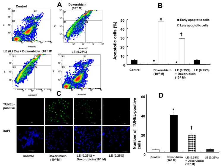
Effects of Intralipid® (lipid emulsion, LE) on the doxorubicin-induced apoptosis of H9c2 rat cardiomyoblasts. The cells were pretreated with doxorubicin (10−5 M) alone for 6 h, LE (0.25%) for 1 h followed by doxorubicin (10−5 M) for 6 h, or LE (0.25%) alone for 7 h. (A) Annexin-V-FITC/propidium iodide staining followed by flow cytometric analysis. (B) Plot (N = 3) showing both early- and late-stage apoptosis after treatment. (C) TUNEL assay showing DNA damage after treatment as detected by immunofluorescence. Nuclei were stained with 4′,6-diamidino-2-phenylindole (DAPI) and are shown in blue. TUNEL-positive cells appear green. Scale bar: 100 μm. (D) Representative plot (N = 3) showing TUNEL-positive cells in different treatment groups. (B,D): Data are shown as the mean ± SD. N indicates the number of independent experiments. * p < 0.001 versus control. † p < 0.001 versus doxorubicin alone.

