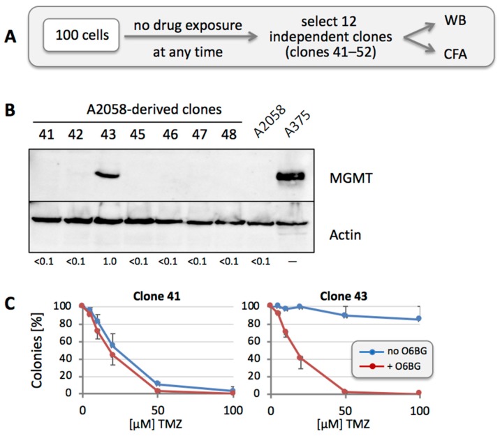
Figure 5.
MGMT protein levels in clones derived from untreated A2058 cells. (A) Treatment schedule was as follows: 100 A2058 cells were seeded into a 10-cm dish. After colonies had formed (in the absence of any drug treatment), 12 of them (numbered 41 through 52) were isolated and expanded for further analysis by WB and CFA. (B) Cell lysates were prepared from all clones and analyzed by WB for MGMT protein levels. Actin was used as a loading control. A lysate of A375 cells was used as a positive control for MGMT protein, and a lysate of parental A2058 cells was used as the negative control. Except for clone 43, all clones were negative for MGMT protein (clones 49 to 52 were negative as well, but not included here; clone 44 was lost). Numbers under the blot indicate quantification of MGMT bands, with reference to the actin signal, and relative to Clone 43 (set at 1.0); <0.1 indicates below detection limit. (C) To confirm drug sensitivity in correlation with MGMT levels, clone 41 and 43 were treated with increasing concentrations of TMZ in the presence or absence of O6BG, and CFA was performed 12 days later (n = 3, ±SE).

