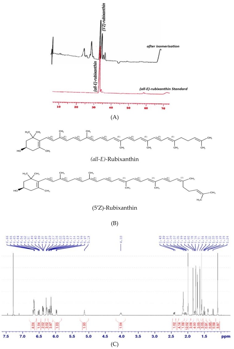
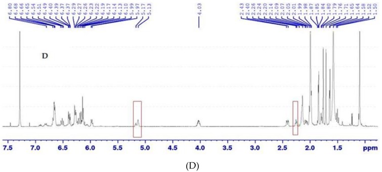
Figure 3.
(A) Chromatographic separation of (all-E)-rubixanthin and (5′Z)-rubixanthin, using a C30 reversed phase column (see text for chromatographic conditions). (B) Structures of (all-E)-rubixanthin and (5′Z)-rubixanthin. (C) 1H nuclear magnetic resonance (NMR) spectrum (600 MHz, 293 K, CDCl3) of (all-E)-rubixanthin prior to light exposition. (D) 1H NMR spectrum (600 MHz, 293 K, CDCl3) of (5′Z)-rubixanthin after light exposition.


