Figure 1.
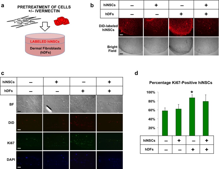
Treatment of dermal fibroblasts with ivermectin induces proliferation in adjacent neural stem cells in 3D co-cultures. (a) Schematic diagram of experimental design. Human dermal fibroblasts (hDFs) and human induced neural stem cells (hiNSCs) fluorescently labeled with DiD dye were separately treated with or without 1 μM ivermectin (as indicated by “+” or “–”, respectively) and subsequently washed repeatedly to remove the drug, seeded into 3D bilayer collagen gel constructs, and cultured for 5 days. (b) Low-magnification view of 3D collagen gel constructs, scale bar: 500 μM. (c) Cryosections of collagen gels immunostained for proliferation marker, Ki67, scale bar: 100 μM. (d) Quantification of Ki67-positive DiD-labeled neural stem cells. *P ≤ 0.05, **P ≤ 0.01, ***P ≤ 0.001; as determined by one-way analysis of variance (ANOVA) with post-hoc Tukey test. Error bars show mean ± SD.

