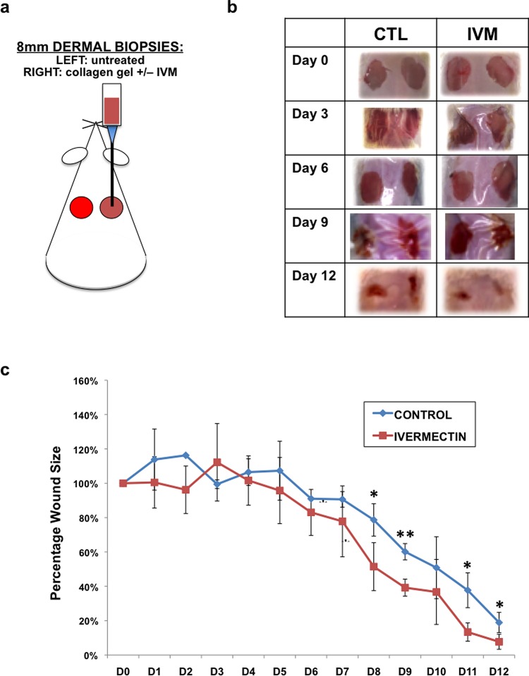
Figure 5.
Ivermectin promotes wound healing of dermal biopsies in vivo. (a) Schematic diagram of experimental design. Biopsies (2 × 8 mm2) were taken from the dorsal dermal layer of each mouse. In the right side wound, 30 μL collagen gels containing 10 μM ivermectin or DMSO (control) were pipetted onto the wound and allowed to solidify. The left side wounds remained untreated, and served as additional controls. Both wounds were sealed using Tegaderm, and wound progression was followed over the course of 12 days. (b) Images of gross morphology of wound healing over time. (c) Quantification of wound size over time. *P ≤ 0.05, **P ≤ 0.01; as determined by two-tailed t-test. Error bars show mean ± SD.

