Figure 4.
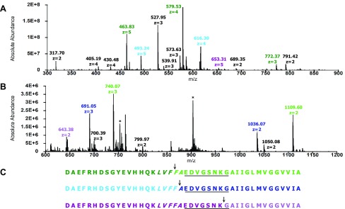
Initial cleavage sites in Aβ42 resulting from IDE-dependent degradation in the presence of resveratrol. (A) Mass spectrum of the primary N-terminal fragments (colored accordingly), which have retention times of 25–30 min. Peaks labeled in black correspond to secondary fragments. (B) Mass spectrum of the primary C-terminal fragments (colored accordingly), which have retention times of 40–43 min. Peaks labeled with asterisks correspond to Aβ42. (C) Peptide maps of Aβ42 depicting initial cleavages at the peptide bonds between Phe19 and Phe20, between Phe20 and Ala21, and between Lys28 and Gly29, corresponding to the color-coded fragments in (A) and (B). Residues in the CHC and loop region are italicized and underlined, respectively.

