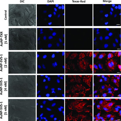
Figure 3.
SVS-1-mediated intracellular delivery of AuNP-conjugates into HeLa cells. Shown are the (DIC), DAPI (nuclei), Texas-Red and merged DAPI (nuclei) and Texas-Red epifluorescence images of control cells and cells incubated with varying concentrations of nanoconjugates of AuNP-TXR (5 nM) and AuNP-SVS-1 (2–5 nM) for 1 h at 37 °C. The Texas-Red dye coupled to AuNP-conjugates allowed visualization of the Au nanocrystal distribution inside the cells. Scale bar ∼ 10 μm.

