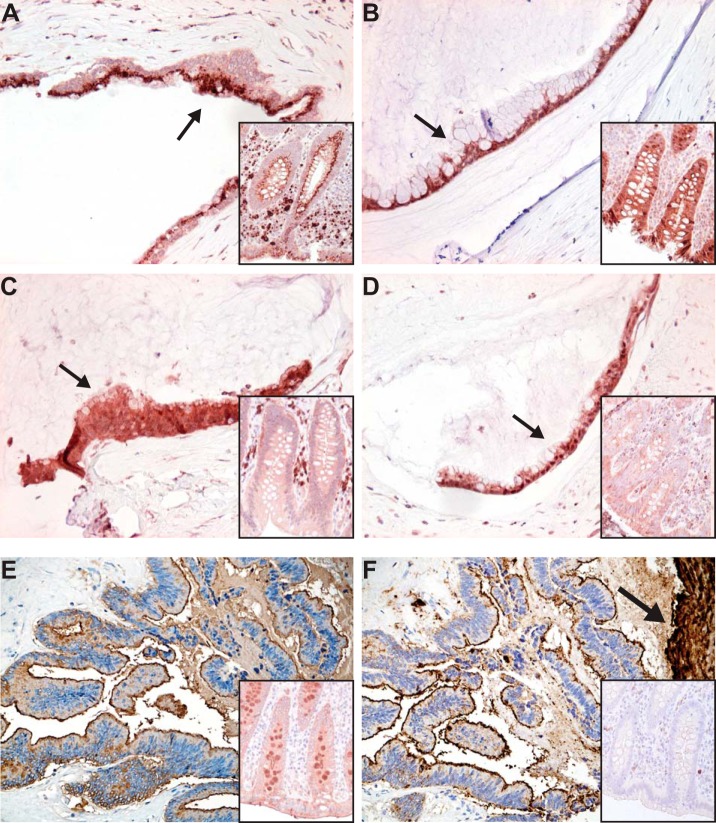
Fig. 5.
Expression of fucosylation-related enzymes and fucosylated glycans in pseudomyxoma peritonei as analyzed by immunohistochemistry and lectin histochemistry. Cytoplasmic expression in tumor cells is shown in Fig. (A) for FUT8, (B) for GMDS, (C) for GMPPA, and (D) for TSTA3 protein. Expression of core fucosylated glycans (E) detected by lectin histochemistry utilizing AAL that preferentially binds to α1,6-linked core fucose residues attached by FUT8 enzyme, and complex fucosylation containing glycan sialyl Lewis x (F) detected by immunohistochemistry. Staining of a control appendix is shown in the insert of each figure. Note the intense staining of mucus in (F) marked by an arrow. Original magnifications, 200X, DAB was used as a chromogen.

