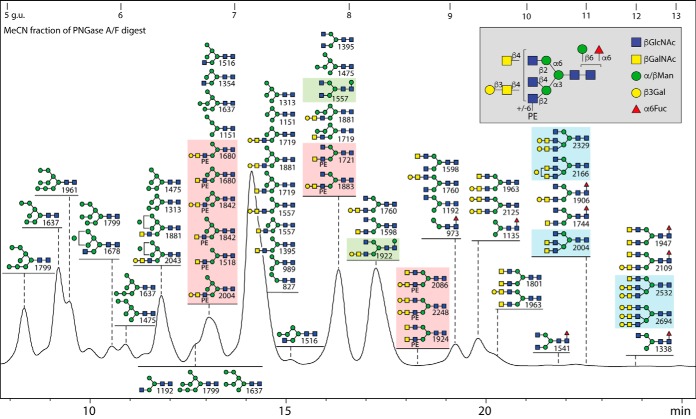
Fig. 1.
RP-HPLC fractionation of the neutral N-glycans from royal jelly. Glycans were released by PNGase F and A before solid phase extraction (to separate the neutral and anionic glycans), pyridylamination and finally fractionation on an RP-amide column (refer to supplemental Fig. S1 for overall MALDI-TOF MS profiles). The chromatogram is annotated with glycan structures (each with the positive mode m/z value for the protonated ion in order of abundance for each fraction) according to the Symbolic Nomenclature for Glycans (circles, galactose or mannose; squares, GalNAc or GlcNAc; triangles, fucose; PE, phosphoethanolamine; see also inset for symbols and linkage information). The HPLC column was calibrated in terms of glucose units (g.u.). Oligomannosidic glycans are annotated by comparison to previous studies on insect glycomes using the same column (unusual isomers were also subject to specific α-mannosidase digestion), whereas other glycans are defined based on MS/MS and digest data. The PNGase F alone and PNGase Ar (after F) digests of the second preparation yielded qualitatively similar chromatograms, but Man9GlcNAc2 was more dominant than Man5GlcNAc2 (see also supplemental Fig. S1A and S1B). The hymenoptera-specific zwitterionic, β-mannosylated and triantennary glycans are highlighted in red, green or blue boxes; the presence of phosphoethanolamine (rather than phosphorylcholine) and of β-mannose, but the relative lack of fucose in this specific glycome, contrasts to the larval N-glycomes of lepidopteran species (19).

