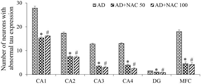
Figure 6.
Quantitative estimation of neurofibrillary tangles (tau positive neurons) in the sub-regions of the hippocampus and prefrontal cortex. In CA1, CA2, CA3. & CA4 regions, a length of 300 µm was selected for quantification; in dentate gyrus (DG) and Media prefrontal cortex (MFC), a 300 µ2 area was selected. Note that in all regions, the number of tau positive neurons significantly decreased in AD + NAC treated groups compared to AD group. Values are expressed as mean ± SE. AD vs. AD + NAC 50: *, p < 0.001; AD vs. AD + NAC 100: #, p < 0.001 (One-way ANOVA, Bonferroni’s multiple comparison test, n = 6 in all groups).

