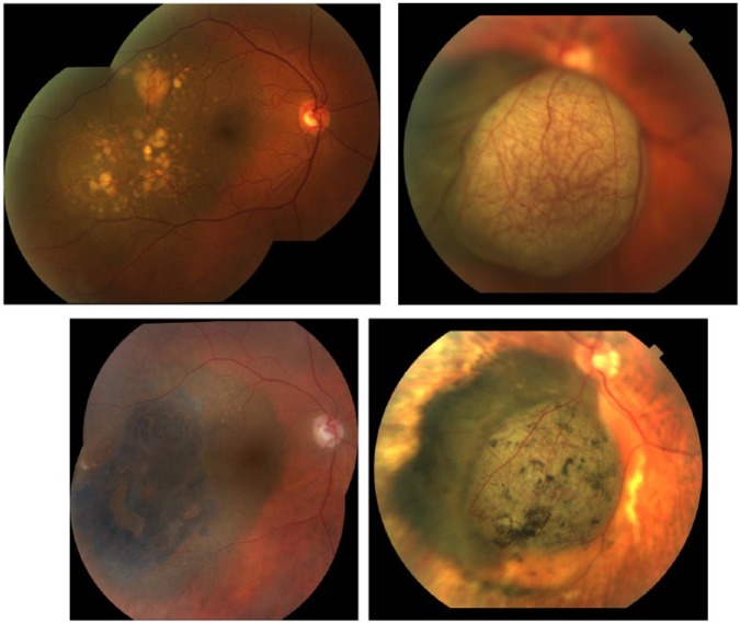
Figure 1.
Fundus photographs of a T2-sized choroidal melanoma (top left) before surgery and (bottom left) 8 years after brachytherapy. Note the change in pigmentation to bluish black, resolution of DRPEDs, and increase in orange pigment lipofuscin. Fundus photographs of a mushroom-shaped choroidal melanoma (top right) before surgery and (bottom right) 3 years after brachytherapy. Note the resolution of intrinsic tumor vascularity.

